Skip to content

Piezosurgery in Jacksonville

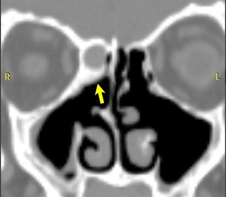
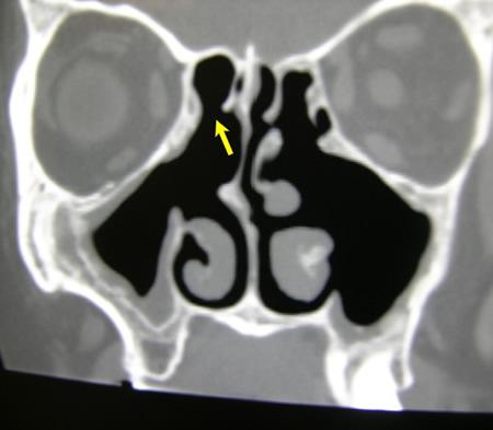
The bone that makes up our sinuses is relatively thin, however, sinus disease can cause it to thicken. Abnormal thick bone may need to be removed in surgery with a small drill. When removing abnormal bone, care must be taken to preserve normal bone that serves as a protective barrier to the neighboring eye and brain structures. Despite improvements in drill technology, risks and complications persist when using drills in sinus surgery due to the close proximity of the sinuses to the eye and brain structures.

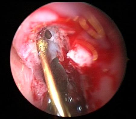
A relatively new technology helps address this challenge by generating sound waves and small vibrations to remove bone.1,2 This technology is called piezosurgery or ultrasonic bone aspiration, and can remove hard bone with small fast vibrations without cutting neighboring softer tissue such as eye, brain or sinus lining membrane, as can happen with the more aggressive rotating action of a drill bit.

If you require sinus surgery and you have thick abnormal bone near a critical structure, our sinus specialists can discuss using this technology for you.

Figure 1
Figure 1a – CT scan of a patient with sinusitis, eye discomfort and headaches. The patient had several previous operations and significant scarring in the sinus. The arrow points to an area of thickened bone and scar that is blocking ethmoid sinus drainage. The white area above the arrow is bone, while the gray area above this represents trapped mucus and infection under pressure.
Figure 1b Endoscopic photo showing the piezosurgery device (gold) treating the thick bone. The device removes bone but preserves the important sinus membrane. The membrane can be seen above the piezosurgery device; it has been opened to drain the infection.
Figure 1b – Endoscopic photo showing the piezosurgery device (gold) treating the thick bone. The device removes bone but preserves the important sinus membrane. The membrane can be seen above the piezosurgery device; it has been opened to drain the infection.
Figure 1c - CT scan of patient in Figure 1a following treatment with the piezosurgery device. The bone, scar tissue, mucus and infection have been removed and normal sinus drainage has been restored to the area. The black area on the CT scan that has replaced the previous gray area, indicates good sinus ventilation. The patient's eye discomfort and headaches improved greatly following surgery.
Figure 1c – CT scan of patient in Figure 1a following treatment with the piezosurgery device. The bone, scar tissue, mucus and infection have been removed and normal sinus drainage has been restored to the area. The black area on the CT scan that has replaced the previous gray area, indicates good sinus ventilation. The patient’s eye discomfort and headaches improved greatly following surgery.

References

1. Vercellotti T, De Paoli S, Nevins M. The piezoelectric bony window osteotomy and sinus membrane elevation: introduction of a new technique for simplification of the sinus augmentation procedure. Int J Periodontics Restorative Dent 2001; 21:561-7.

2. Vercellotti T, Pollack AS. A new bone surgery device: sinus grafting and periodontal surgery. Compend Contin Educ Dent 2006; 27:319-25.

Request an Appointment Today!

Come be a part of the North Florida Sinus Center family. Request an appointment with one of our board-certified providers today!